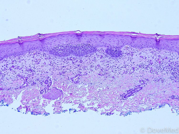
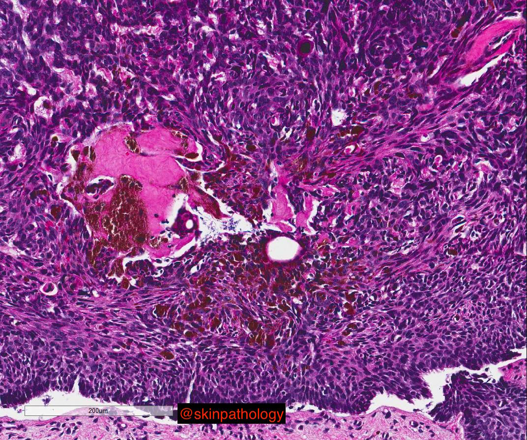
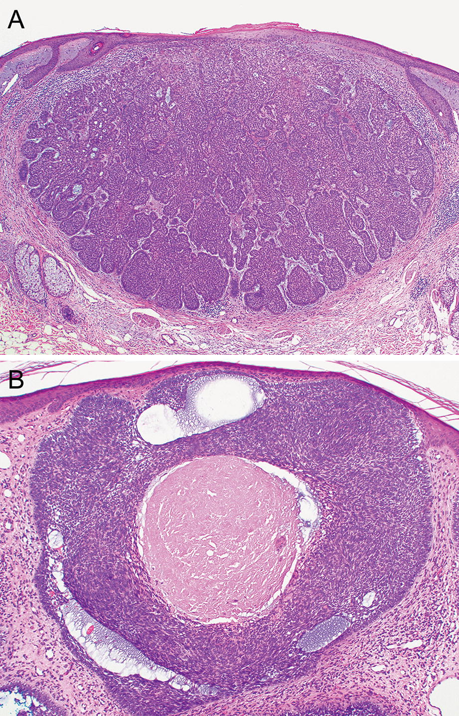
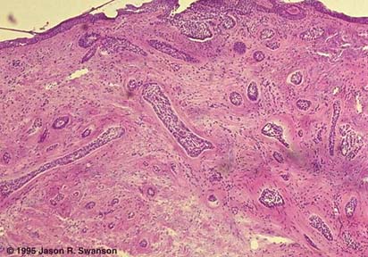
Nodular Basal Cell Carcinoma Histology
Bcc is also known as rodent ulcer and basalioma.

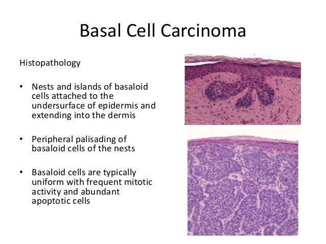
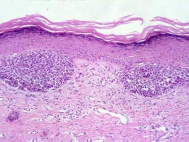
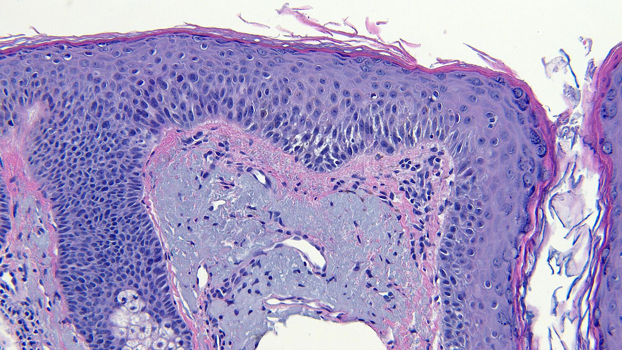
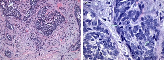
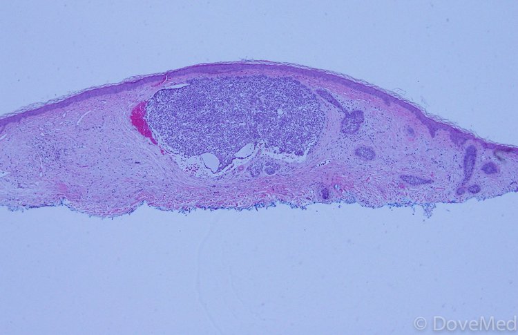
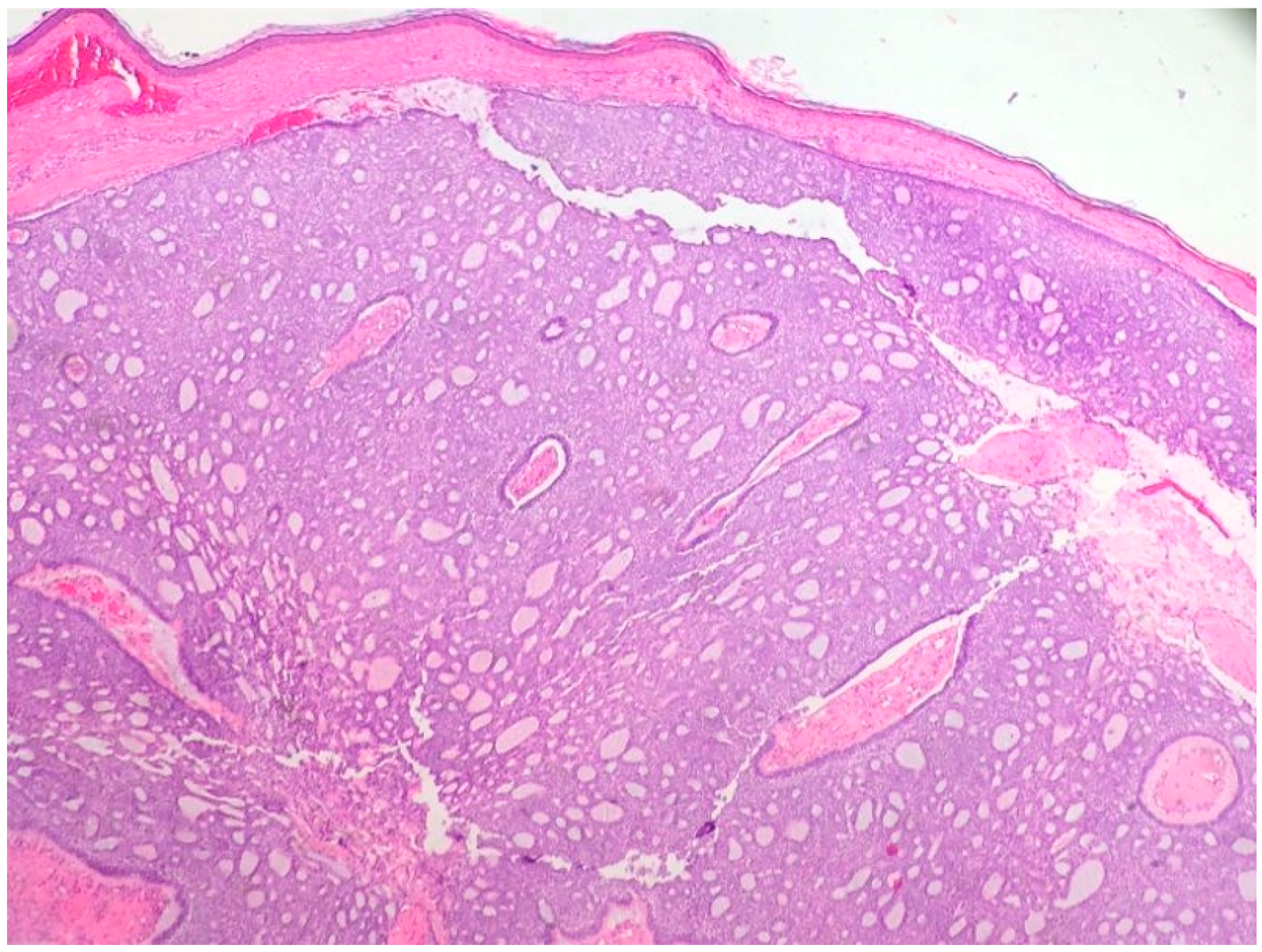
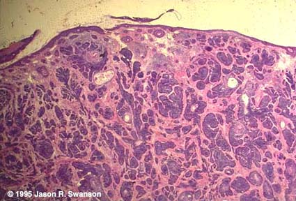
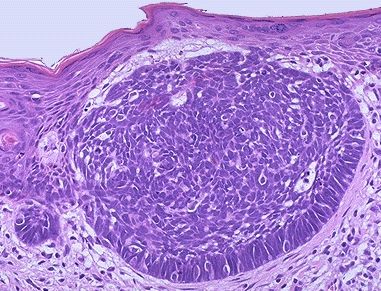
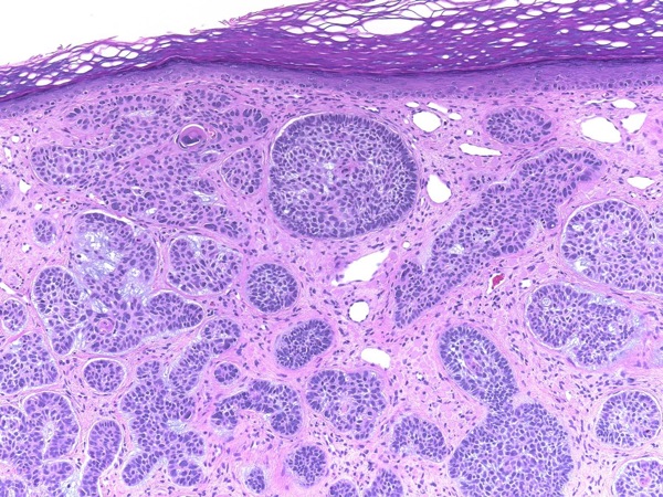
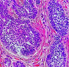
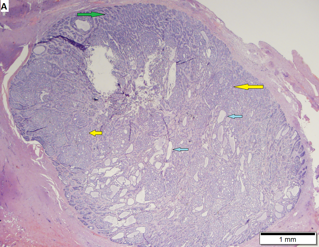
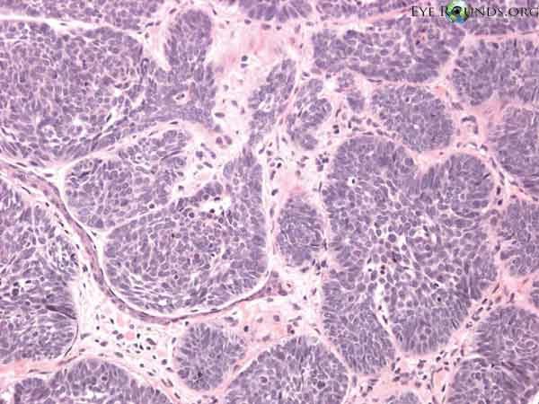
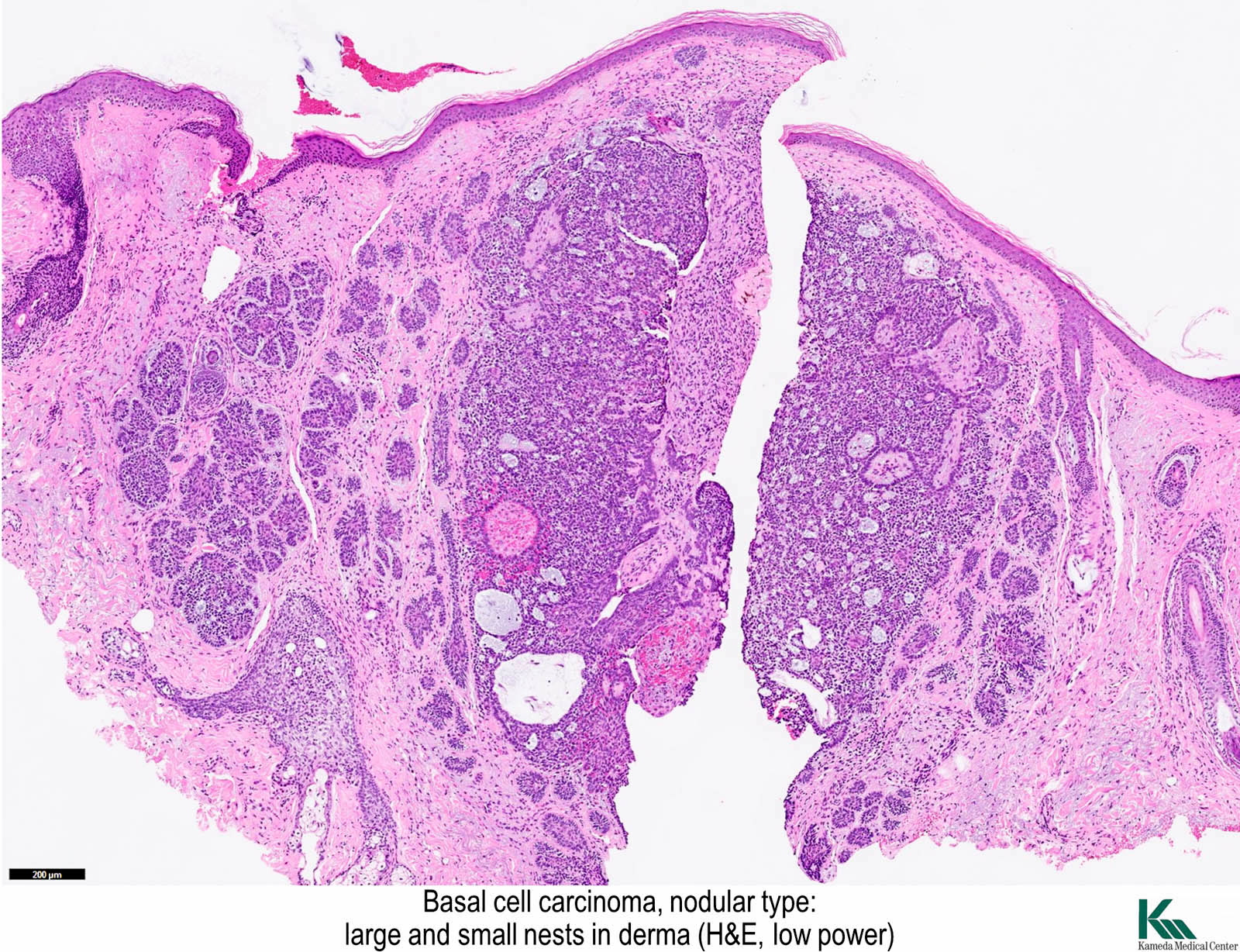
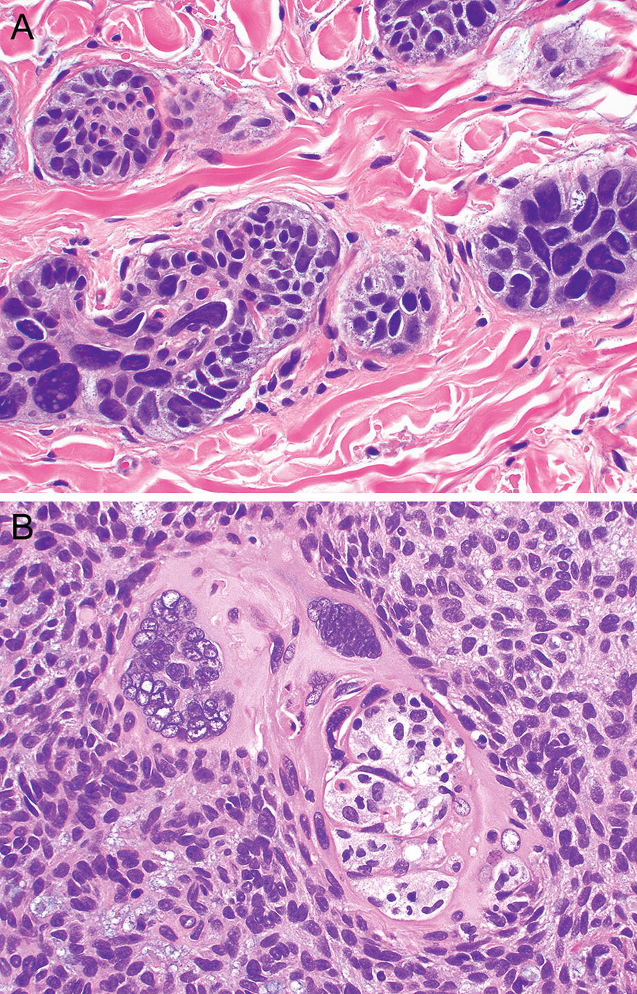
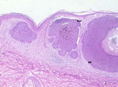
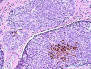
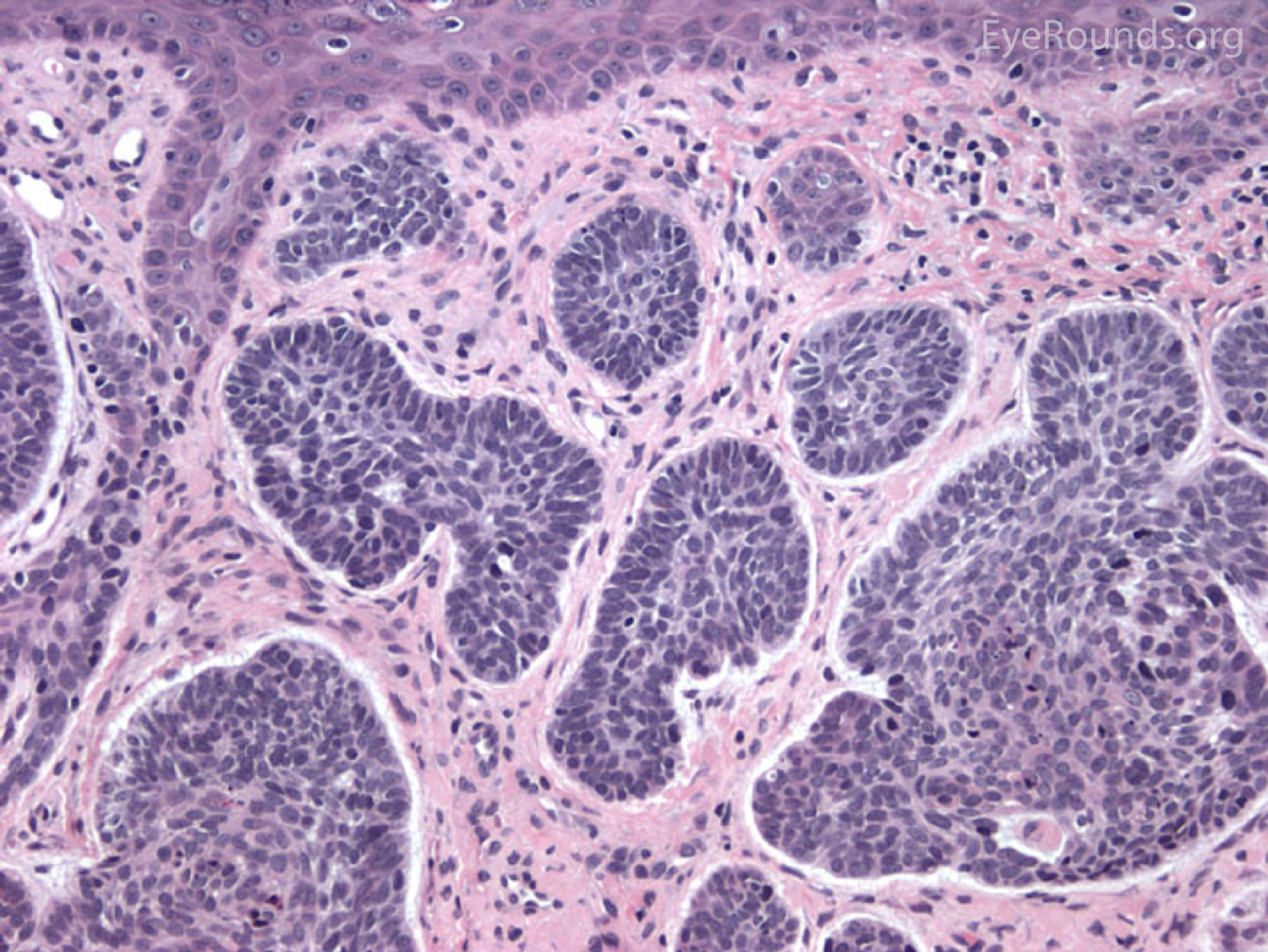
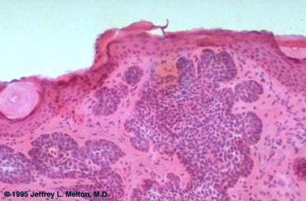
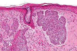
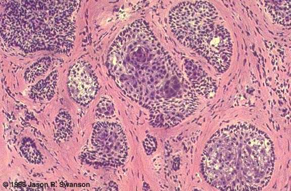
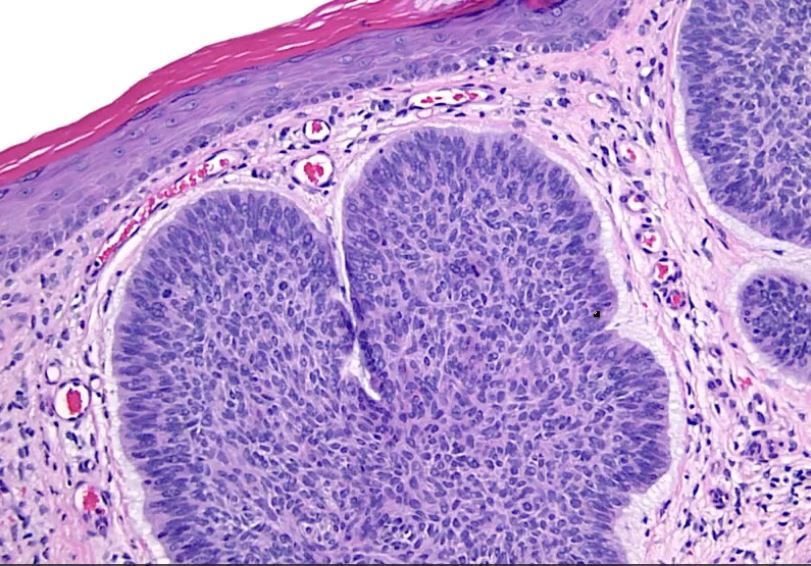
Nodular basal cell carcinoma histology. The groups of basaloid cells have peripheral palisading of the nuclei and are surrounded by a myxoid stroma. High power microscopy shows. Programmed cell death physiology and pathology. Basal cell carcinoma bcc is a common locally invasive keratinocyte cancer also known as nonmelanoma cancer.
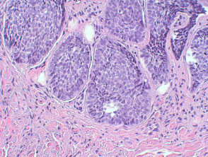
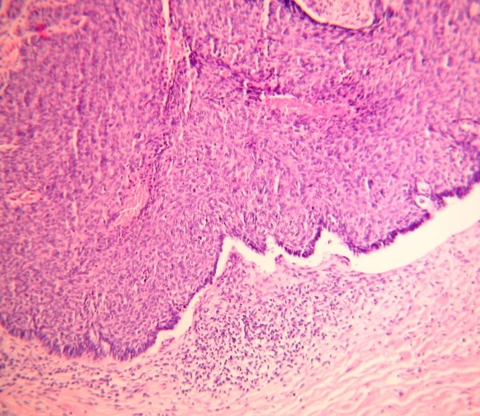
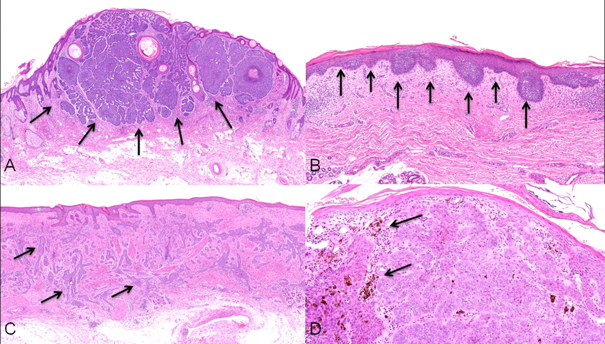
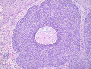
The groups of basaloid cells are well demarcated from the stroma and show focal clefting from the stroma. The metatypical bcc is an aggressive growth neoplasm arising in sun damaged skin and showing irregular tongues of tumor andor an admixed nodular bcc component a. It is the most common form of skin cancer. Larger tumors also have a greater tendency to recur after treatment.
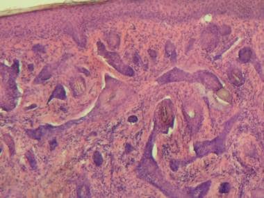
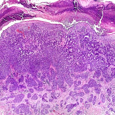
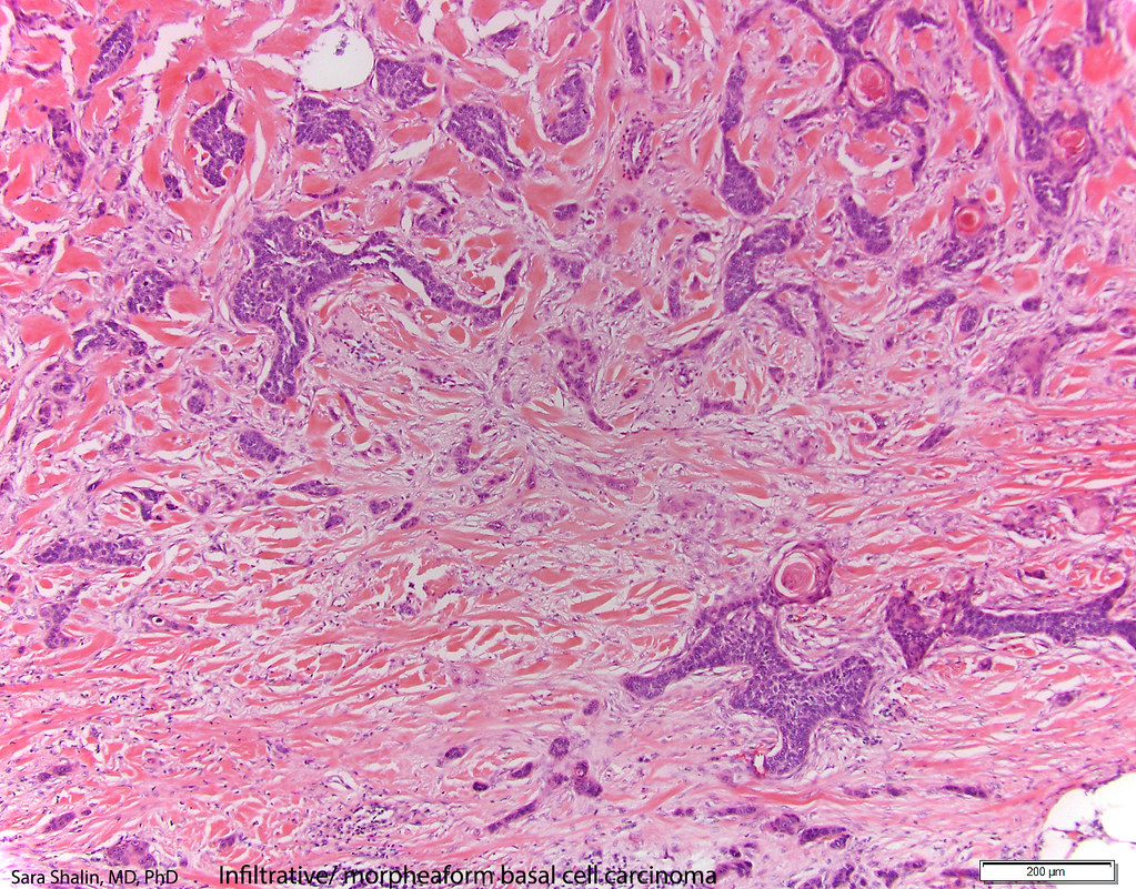
They can also infiltrate into the adjoining soft tissues and nerves. An unreflective line is seen bordering the tumor islands white arrows corresponding to mucinous cleftingperipheral palisading often recognized in histology sections. Nodular basal cell carcinoma of skin is the most common type of bcc that is present as nodules on the skin usually in the head and neck area some nodules may grow to large sizes and ulcerate. Histopathology of skin basal cell carcinoma.
Patients with bcc often develop multiple primary tumours over time. Histology of basal cell carcinoma. The edge of the tissue focally has basal cell carcinoma the deep aspect is clear. Nodular basal cell carcinoma located on the cheek showing disruption of normal layering and hyporeflective oval structures white asterisks corresponding to tumor islands.
The basaloid epithelium typically forms a palisade with a cleft forming from the adjacent tumour stroma figure 2.

